Context
We aim at developing methods allowing to create subject-specific computational models of the mitral valve. We are working on automatically extracting the valve geometry as well as building a physically-based simulation of the valve closure. Building bio-mechanical models of physiological behavior is difficult due to complex structures of deformable anatomical parts.
Collaborations
This work is in collaboration with Harvard University through the Inria associate research team CURATIVE ![]() .
.
Results
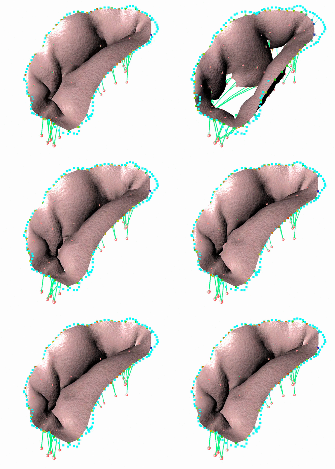
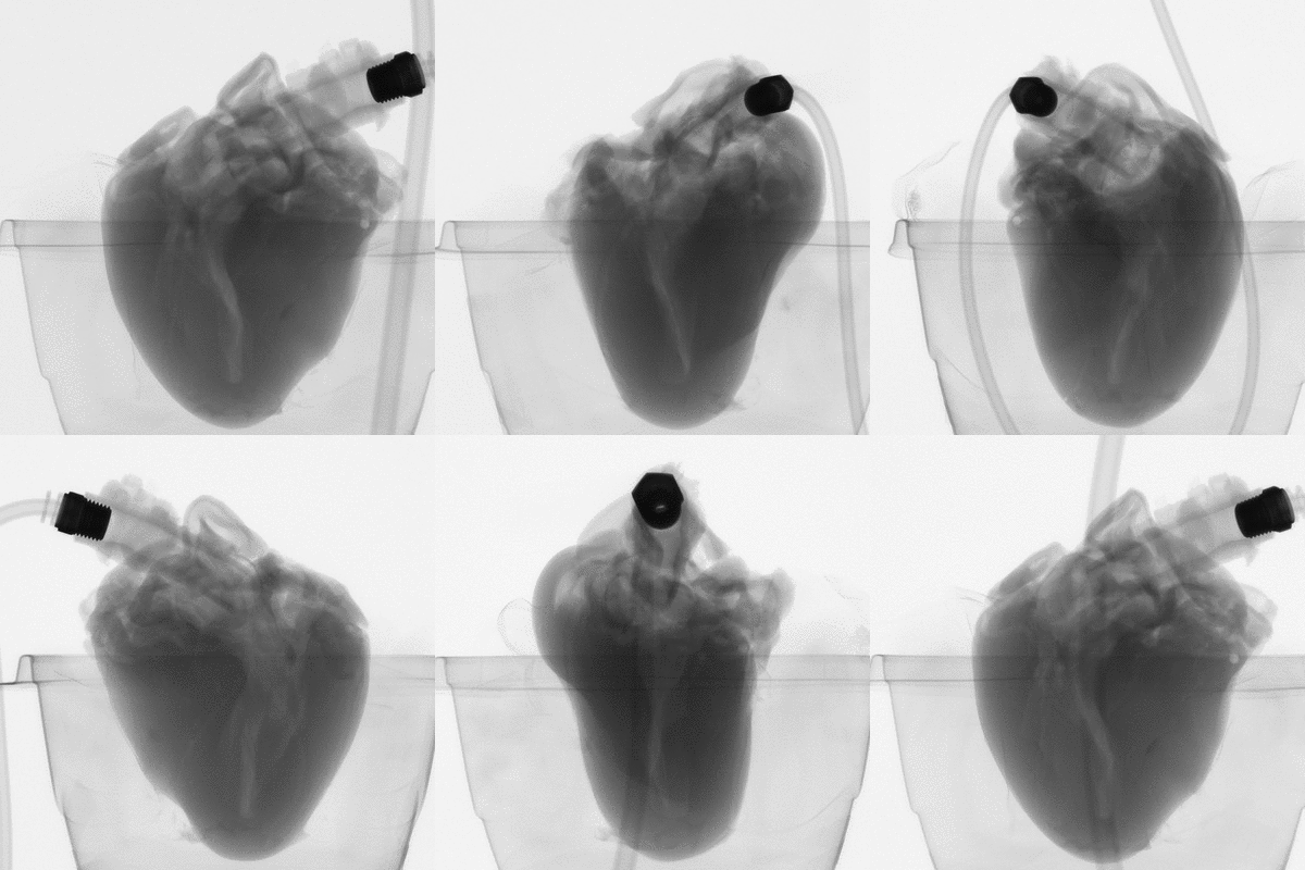
We have proposed a method to semi-automatically extract structures and boundary conditions of such a structure from micro CT scans [1]. Anatomical parts were extracted based on a study of the orientation of the model skeletization line segments. Boundary conditions were found by analyzing the topology between anatomical parts. A fast FEM model [2] developed with SOFA (Simulation Open Framework Architecture) was then carried out to reproduce leaflets closing during systolic peak based on the previous study. An implicit Euler was used as the integration scheme to allows the simulation to remain stable while using large time steps and then be interactive. All the valve components have their own mechanical model. Results on explanted porcine heart showed that it is possible to predict the shape of the leaflets with an average surface error of 1mm.

microCT scan images before 3D reconstruction


